Appearance
Saccular lobes
A saccular aneurysm is a dead end of the vessel: a sac that bulges from the wall and leads nowhere. When it grows at a junction, as a terminal aneurysm at the end of the internal carotid artery does, it is part of no branch. The sections of the branches around it cut through it, and the extension of the parent behind the apex covers only part of it. A tube cannot describe it, whatever the shape of its sections. furca models such a sac as a lobe: a closed surface star-shaped about a centre, joined to the branches by the same smooth union as the branches are joined to each other.
Loading the model…
Drag to rotate, scroll or pinch to zoom, right-drag to pan
A growing terminal aneurysm of the left internal carotid artery (data/vmr/cerebral-aneurysm-branched/0207_H_CERE_CA), its wall coloured by the distance to the model (0 to 10% of the radius of the carotid). Switch the layers: without lobes no branch follows the sac, and the error reaches half the radius; with a lobe it stays below 2%.
The model
A lobe is the region lobes.degree, 12), orthonormal on the sphere:
Its implicit function, in units of its mean radius
negative inside and zero on its wall. It enters the smooth union of the vessel as one more term, present everywhere (
Spherical harmonics describe closed surfaces star-shaped about a point, as in the spherical-harmonic shape descriptors of Brechbühler, Gerig & Kübler [32]. Most saccular aneurysms are star-shaped about a point of their sac. Here the sac is open at its neck, where it meets the vessel, and there the union with the branches takes over.
Finding the lobes
A lobe is where the flow stagnates. With
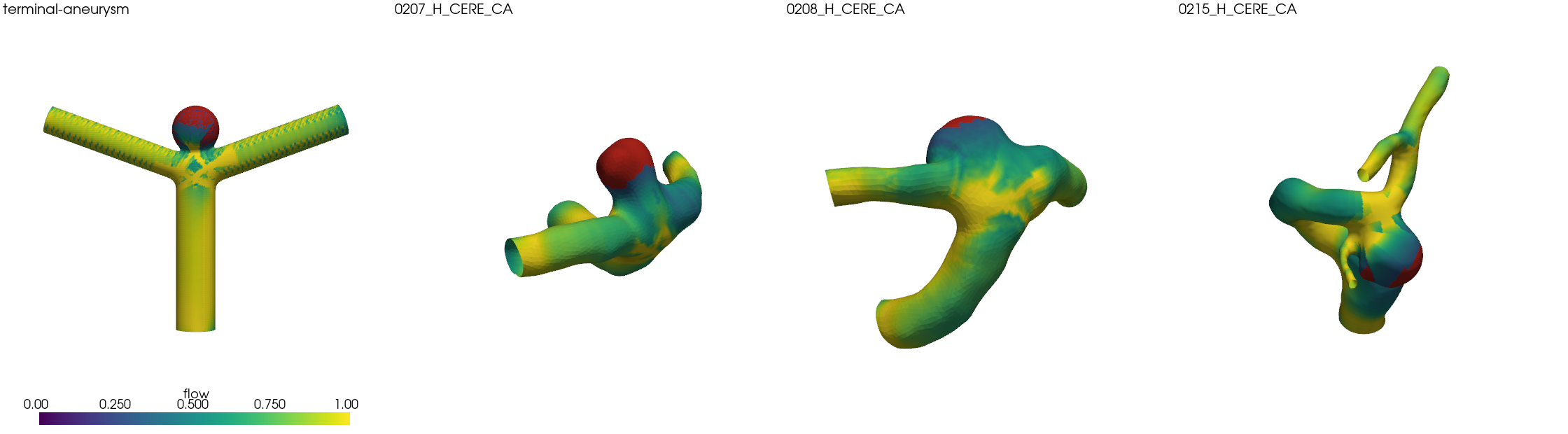
The flow on the wall of the synthetic terminal aneurysm and of three cerebral aneurysms (0207, 0208, 0215); in red where it is below 0.3.
Then:
- Cores. The connected regions where the flow is below
lobes.stagnation(0.3) and whose area is at leastlobes.min_area(one) times, with the mean radius of the inlet and outlets, are the stagnant cores of lobes. Smaller ones, such as the crotch of a junction, are not. - Centre. The centre is that of the sphere that best fits the core, algebraically:
in least squares, weighted by area. A cap of a sphere fixes its centre and radius well. - Region. The lobe spans its core and, grown from it across edges, the wall within
lobes.sphere_tolerance(one fifth) of the sphere's radius from the sphere, as far as the neck. Cores whose regions overlap (a sac whose flow stagnates in more than one patch) make one lobe.
The vertices of a lobe are left out of the fits of the branches and out of their level rings, so that the centerline of a branch does not swerve into the sac.
Fitting a lobe
Every lobe is first fitted alone, to the vertices of its region, by penalized least squares on the distances from its centre along their directions. The penalty is the squared Laplace–Beltrami operator on the unit sphere,
The sac and its morphology
The sac of a lobe is the part of its wall that lies outside every branch: the wall of the aneurysm. Its boundary is the neck, where the sac opens into the vessel. Both are found on the lobe's wall sampled on an icosphere in its frame (20 · 4⁵ triangles): the sac is the largest connected part where the smooth union of the branches alone is positive, and the neck is its rim. The neck has a centre (its centroid by length) and a normal (its area vector, from the vessel into the sac), which give the plane of the neck. As in the aneurysm literature, where the sac is isolated by a cut at its neck, its volume and area are those of the lobe beyond that plane, closed by it. The indices are those of Raghavan, Ma & Harbaugh [33] and Dhar et al. [34]:
| Index | Definition |
|---|---|
| Height | Largest distance from the plane of the neck to the sac. |
| Max height | Largest distance from the centre of the neck to the sac. |
| Max diameter | Largest distance between two points of the sac. |
| Neck diameter | Twice the mean distance of the neck from its centre, in its plane. |
| Parent diameter | Mean diameter of the branches the neck opens into, weighted by the length of neck each dominates. |
| Aspect ratio | |
| Size ratio | |
| Volume | Of the sac beyond the plane of its neck (the area without the plane). |
| Nonsphericity | |
| Undulation | |
| Ellipticity |
They are in the summary of every encoding (furca encode, furca inspect, the application and Vessel.summary() in Python), and Vessel.sac(j) returns them with the wall and the neck of the sac. Because they are computed from the model, they are as smooth as the lobe: Vessel.with_lobe_degree(l) gives the vessel with its lobes described up to degree
On the synthetic terminal aneurysm, a sphere whose cap below the neck lies inside the vessel, the height, volume, area and largest diameter of the sac are those of the sphere without that cap within 2–3%, and its undulation is below 0.01. On the four cerebral aneurysms, at degrees 4, 8 and 12 for the aspect ratio:
| Case | H (mm) | N (mm) | AR, degree 4 / 8 / 12 | SR | V (mm³) | NSI | UI | EI |
|---|---|---|---|---|---|---|---|---|
0207_H_CERE_CA | 3.48 | 3.15 | 1.05 / 1.07 / 1.10 | 1.32 | 25.2 | 0.151 | 0.056 | 0.277 |
0208_H_CERE_CA | 3.18 | 4.56 | 0.71 / 0.72 / 0.70 | 1.27 | 39.4 | 0.043 | 0.012 | 0.328 |
0215_H_CERE_CA | 7.93 | 4.65 | 1.54 / 1.52 / 1.70 | 2.50 | 208.7 | 0.172 | 0.051 | 0.250 |
0216_H_CERE_CA | 2.86 | 3.23 | 1.03 / 0.87 / 0.89 | 0.95 | 18.0 | 0.135 | 0.070 | 0.288 |
In both matched pairs the aneurysm that grew (0207, 0215) has a larger aspect ratio and a larger nonsphericity than the one that stayed stable (0208, 0216), as the literature on rupture and growth reports; two pairs show the indices at work, not a finding. The aspect ratio changes by a few percent between degrees 4 and 12, except in 0215, where the neck lies next to the mouth of the posterior communicating artery and moves with the detail of the lobe there.
Validation
On the synthetic terminal aneurysm (furca synth terminal-aneurysm: a T-shaped bifurcation with a sphere on its apex) and on four cerebral aneurysms in branched vessels from the Vascular Model Repository: two of the terminus of the internal carotid artery and two of the posterior communicating artery, each a matched pair of one that grew and one that stayed stable. The table gives the distance from the open wall to the model, relative to the radius of the parent vessel. For the synthetic case it is the distance from the model to the exact wall, away from the cut ends.
| Case | Branches | Lobes | Without lobes: mean / max | With lobes: mean / p99 / max | Time |
|---|---|---|---|---|---|
terminal-aneurysm | 3 | 1 | 4.7 s | ||
0207_H_CERE_CA | 2 | 1 | 2.14% / 59.84% | 0.11% / 0.70% / 1.55% | 1.8 s |
0208_H_CERE_CA | 2 | 1 | 0.23% / 2.79% | 0.16% / 1.02% / 2.70% | 0.6 s |
0215_H_CERE_CA | 3 | 1 | 0.24% / 8.83% | 0.21% / 1.48% / 7.55% | 4.6 s |
0216_H_CERE_CA | 3 | 1 | 0.20% / 11.02% | 0.09% / 0.90% / 3.61% | 3.1 s |
The lobes change nothing where there is no sac: every other vessel of Validation, synthetic or real, has none, and its encoding is the same. In the terminal aneurysms the error falls from half the radius to a few percent: the sac of 0207 is fitted to 1.6% at most. In 0208 the sac is small and was already covered by the branches, and the lobe changes little. In 0216 the largest error falls from 11% to 3.6%. In 0215 the largest error, 7.5%, is a crease next to the mouth of the posterior communicating artery, which leaves right beside the sac, and the 99th percentile falls from 2.2% to 1.5%. In the synthetic case the largest error, 4.5%, is at the neck, where the fillet of the synthetic union differs from that of the model; the sac itself is a sphere and is fitted to a fraction of a percent.
Unbranched vessels
A sac on the side of an unbranched vessel is a dead end of the flow too. The tube's general wall follows it as long as every section meets the wall in one closed curve, but not where the sac lies inside a loop of the vessel, as in the carotid siphon, where a section crosses the sac and the vessel twice. An unbranched vessel whose wall has a lobe is therefore encoded both ways: as a tube, and as one branch with its lobes. The lobes are kept when they leave at most lobes.tube_residual (one half) of the tube's RMS residual; otherwise the vessel stays a tube, and its encoding is the same as without lobes.
Loading the model…
Drag to rotate, scroll or pinch to zoom, right-drag to pan
An unbranched cerebral aneurysm whose sac lies inside a tight loop of the carotid siphon (data/vmr/cerebral-aneurysm/0203_H_CERE_CA), its wall coloured by the distance to the model (0 to 10% of the radius). Switch the layers: the general wall of the tube cannot follow the sac where its sections cross it; with a lobe the error stays below 5%.
On the synthetic side aneurysm (furca synth side-aneurysm: a straight tube with a sphere on its side), the lobe leaves a fifth of the tube's RMS residual and its largest error is 2.5% of the radius, against 15% for the tube. On the two unbranched cerebral aneurysms (Cerebral aneurysms), the lobe of 0203 leaves 8% of the tube's RMS residual and is kept: the mean error falls from 2.7% to 0.6% of the radius, the largest from 38% to 4.4%. The sac of 0204 has blebs of its own, which a surface star-shaped about one centre does not follow as well as the general wall of the tube: its lobe leaves 1.25 times the tube's residual, and the tube is kept. Synthetic tubes, aortas and aortic arches have no lobes.
Limitations
- Lobulated sacs. A sac with blebs or lobes of its own is described up to the degree of its harmonics, and its region stops growing where it departs from the sphere of its core. In an unbranched vessel, the tube's general wall may then describe it better, and is kept (as in
0204). - Cohorts need the same number of lobes in every subject.
- The neck is where the model's lobe leaves its branches, and its plane is fitted to it: where a branch leaves right beside the sac, as in
0215, it carries the uncertainty of the wall there into the height and the aspect ratio.